Bursitis Of The Foot Pain Treatment
Overview
A bursa is a fluid-filled sac. There are hundreds of deep and superficial bursae throughout the human body. They are typically located near major joints. Bursae are situated between bone and soft tissues like tendons, ligaments, muscles, and skin. They serve as a cushioning pad to absorb shock. The fluid within the sac is secreted to assist with friction-free movement as the soft tissues move across a bony area.
Causes
As ligaments and tendons stretch and tear, blood from ruptured blood vessels becomes trapped in the local tissues. As the trapped blood clots up, it sticks the tissues together creating adhesions. Adhesions cause pain, inflammation and restricted movement because the layers of tissue that used to slide smoothly across one another now adhere and snap which interferes with normal functioning. It is essential to break up clotted blood as quickly as possible to prevent adhesions and scar tissue from forming.
Symptoms
Posterior heel pain is the chief complaint in individuals with calcaneal bursitis. Patients may report limping caused by the posterior heel pain. Some individuals may also report an obvious swelling (eg, a pump bump, a term that presumably comes from the swelling's association with high-heeled shoes or pumps). The condition may be unilateral or bilateral. Symptoms are often worse when the patient first begins an activity after rest.
Diagnosis
Bursitis is usually diagnosed after a careful physical examination and a full review of your medical history. If you garden and spend a lot of time on your knees, or if you have rheumatoid arthritis, tell your doctor, this information can be very helpful. During the physical exam, he or she will press on different spots around the joint that hurts. The goal is to locate the specific bursa that is causing the problem. The doctor will also test your range of motion in the affected joint. Other tests usually aren?t required to diagnose bursitis, but your doctor may suggest an MRI, X-ray or ultrasound to rule out other potential causes of pain.
Non Surgical Treatment
So what can you do to alleviate this type of pain in the foot? If the bursitis pain is occurring on the toes, bunion or back of the heel area the smart money would be on eliminating the shoes that seem to aggravate the condition. Eliminating these shoes may not in itself clear up the problem but you can be sure that if you continue to wear the offending shoes nothing you or your doctor do will permanently ?fix? the problem. A recurring theme that I use throughout this site that if you put an abnormally shaped foot in a dressy shoe it is literally the same as trying to put a square peg in a round hole, it will not fit. OK, so you threw away those dressy shoes and the foot still hurts, now what? Depending on the severity of the pain, over the counter anti-inflammatory medication may do the trick. The key here is to take the medication on an ongoing basis, according to the directions on the package to build up therapeutic blood levels. Assuming you can tolerate this type of medication, along with alternative treatments you can try). take the medication for 10-14 days. Stop if the symptoms have not dramatically improved. Icing the area during this period may also help reduce the symptoms.
Surgical Treatment
Surgery. Though rare, particularly challenging cases of retrocalcaneal bursitis might warrant a bursectomy, in which the troublesome bursa is removed from the back of the ankle. Surgery can be effective, but operating on this boney area can cause complications, such as trouble with skin healing at the incision site. In addition to removing the bursa, a doctor may use the surgery to treat another condition associated with the retrocalcaneal bursitis. For example, a surgeon may remove a sliver of bone from the back of the heel to alter foot mechanics and reduce future friction. Any bone spurs located where the Achilles attaches to the heel may also be removed. Regardless of the conservative treatment that is provided, it is important to wait until all pain and swelling around the back of the heel is gone before resuming activities. This may take several weeks. Once symptoms are gone, a patient may make a gradual return to his or her activity level before their bursitis symptoms began. Returning to activities that cause friction or stress on the bursa before it is healed will likely cause bursitis symptoms to flare up again.
Flexible Hammertoes Treatment
 Overview
A hammertoes is a toe that is bent because of a muscle imbalance around the toe joints. The imbalance causes the toe to bend at one or more joints, pushing the middle of the toe upward in a claw-like position. If you notice such changes, it is important to seek proper treatment. Hammer toes never get better without some type of intervention and the sooner it is treated, the better the outcome.
Causes
Footwear can contribute significantly to the development of hammertoes. Shoes that are too small force your toes into a curled position. Over time, your toe tendons adjust to this positioning, causing your toe or toes to hold a hammered shape. Athletes may be especially susceptible, because of the increased forces on the toes from shoes that are too small or tight. Heel elevation in footwear is also problematic, as it causes your toes to be pushed into the shoe?s toe box. Heel elevation additionally contributes to muscle imbalance. A common example of this is when your Achilles tendon-the tendon at the back of your leg that attaches your calf muscles to your heel bone-is too tight, causing the tendons on the top of your foot that attach to your toes to work too hard and hold your toes in an unnatural, elevated position.
 Symptoms
If you have any of these symptoms, do not assume it is due to hammer toe. Talk to your doctor about symptoms such as a toe that curls down, corns on the top of a toe, calluses on the sole of the foot or bottom of the toe, pain in the middle joint of a toe, discomfort on the top of a toe, difficulty finding any shoes that fit comfortably, cramping in a toe, and sometimes also the foot and leg, difficult or painful motion of a toe joint, pain in the ball of the foot or at the base of a toe.
Diagnosis
The treatment options vary with the type and severity of each hammer toe, although identifying the deformity early in its development is important to avoid surgery. Your podiatric physician will examine and X-ray the affected area and recommend a treatment plan specific to your condition.
Non Surgical Treatment
Conservative treatment starts with new shoes that have soft, roomy toe boxes. Shoes should be one-half inch longer than your longest toe. (Note: For many people, the second toe is longer than the big toe.) Avoid wearing tight, narrow, high-heeled shoes. You may also be able to find a shoe with a deep toe box that accommodates the hammer toe. Or, a shoe specialist (Pedorthist) may be able to stretch the toe box so that it bulges out around the toe. Sandals may help, as long as they do not pinch or rub other areas of the foot.
Surgical Treatment
Any surgery must be carefully considered and approached in a serious manner, as any procedure is serious for the patient. But in most cases the procedure is relatively straight forward. The surgery can be done using local hammertoes anesthetic and does not require hospitalization. The patient goes home in a special post-operative shoe or a regular sandal, and in most cases can walk immediately. That's not to say that the patient is walking or functioning normally immediately after the procedure. The patient must take some time off work to rest the foot and allow it to heal.
How To Deal With Bunions
Overview
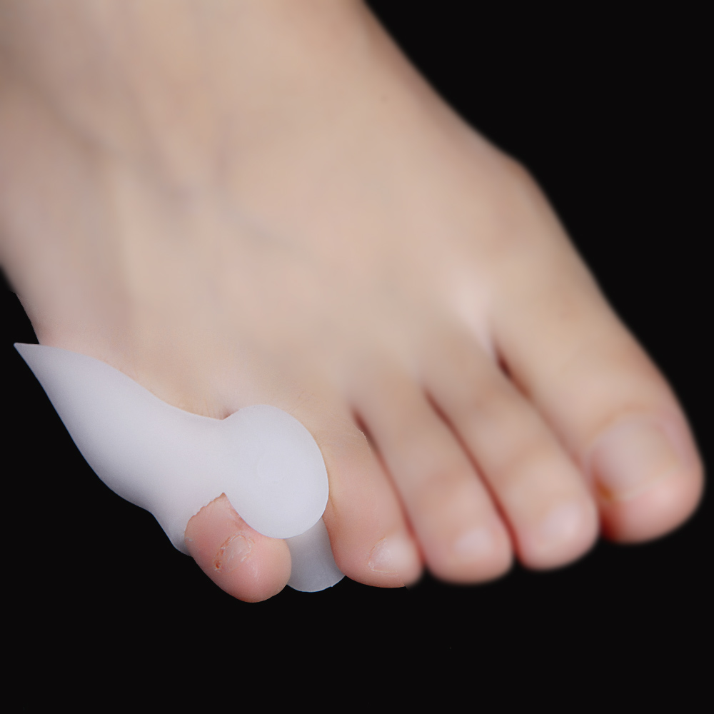
 A Bunion is one of the most common foot ailments which usually occur near the joint at the base of the big toe. It is actually a bony protrusion which consists of excess or misaligned bone in the joint. Although they may develop on the fifth or little toe, bunions usually occur at the base of the big toe. In addition to causing pain, a bunion changes the shape of your foot, making it harder to find shoes that fit. The good news however, is that you don?t have to hobble for the rest of your life, bunions can be treated.
Causes
The causes of bunions are "multifactorial". In other words generally many things contribute to their occurence. Shoewear per se is a contributory factor but it isn't possible to say that the type of shoewear is necessairily relevant. Often there is a hereditary component with other family members also suffering with the same condition.
Symptoms
Pain in the toe joint and surrounding area. Painful to touch or press, and when walking. Growth of a bony lump (exostosis) at the side of the big toe joint. Irritated skin around the bunion. Redness. Thickening of overlying skin. Blisters may form more easily. Deformed bones, joints and ligaments as the big toe shifts towards the other toes. As the big toe shifts, its base becomes more prominent, forming the bunion. Eventually the big toe is forced to lie over, or more commonly under, the second toe. The second toe of patients who have bunions commonly forms a hammer toe. Trouble with shoes. It is difficult to find shoes that fit properly. Bunions may force you to buy a larger size shoe to accommodate the width the bunion creates. Eventually it hurts to wear any shoe, or even walk barefoot.
Diagnosis
Your doctor is very likely to be able to diagnose your bunion simply by examining your foot. Even before that, he or she will probably ask about your family and personal medical history and evaluate the types of shoes you wear. You'll be asked about your symptoms, when they started and when they occur. You may also be asked to flex your toe so that your doctor can get an idea of your range of motion. He or she may order x-rays in order to determine the extent of your deformity.
Non Surgical Treatment
A bunion may only need to be treated if it's severe and causing significant pain and discomfort. The different treatments for bunions are described below. If possible, non-surgical treatment for bunions will be used, which your GP can discuss with you. Non-surgical treatments can ease the pain and discomfort caused by a bunion, but they can't change the shape of your foot or prevent a bunion from getting worse over time. Non-surgical treatments include painkillers, bunion pads, orthotics, wearing suitable footwear, These are discussed in more detail below. If your bunion is painful, over-the-counter painkillers such as paracetamol or ibuprofen may be recommended.Bunion pads may also ease the pain of a bunion. Reusable bunion pads, made of either gel or fleece, are available over the counter from pharmacies. Some are adhesive and stick over the bunion, while others are held against your foot by a small loop that fits over your big toe. Bunion pads stop your foot rubbing on your shoe and relieve the pressure over the enlarged joint at the base of your big toe. Orthotics are placed inside your shoes to help realign the bones of your foot. They may help relieve the pressure on your bunion, which can ease the pain. However, there's little evidence that orthotics are effective in the long term. It's important that the orthotic fits properly, so you may want to seek advice from your GP or podiatrist (a specialist in diagnosing and treating foot conditions), who can suggest the best ones for you. 
Surgical Treatment
There is no "standard" bunion, but rather a complex range of joint, bone, muscle, tendon and ligament abnormalities that can cause variation in each bunion's make-up. As a result, there are a broad variety of surgical techniques for dealing with bunions. Most surgical procedures start with a simple bunionectomy, which involves excision of swollen tissues and removal of the enlarged boney structure. While this may remove the troublesome tissues, however, it may not correct other issues associated with the bunion. The surgeon may also need to tighten or loosen the muscles, tendons and ligaments around the MTP joint. Realign the bone by cutting it and shifting its position (a technique called osteotomy), realigning muscles, tendons and ligaments accordingly. Use screws, wires or plates to hold the joint surfaces together until they heal. Reconstruct a badly damaged joint or replace it with an artificial implant.
The Facts Related To Overpronation Of The Feet
Overview
When the ankles over-pronate, it throws everything out of alignment. The bones in the feet can actually shift over time, and the muscles and tendons in the leg and ankle will twist. This places repetitive stress on the ligaments, tendons, joints and muscles of the foot, which can cause injury. If your feet are over-pronating, you may notice your muscles feet unusually tired when you way or stand, or they may ache. But symptoms will vary by your weight, age and activity.
Causes
Unless there is a severe, acute injury, overpronation develops as a gradual biomechanical distortion. Several factors contribute to developing overpronation, including tibialis posterior weakness, ligament weakness, excess weight, pes planus (flat foot), genu valgum (knock knees), subtalar eversion, or other biomechanical distortions in the foot or ankle. Tibialis posterior weakness is one of the primary factors leading to overpronation. Pronation primarily is controlled by the architecture of the foot and eccentric activation of the tibialis posterior. If the tibialis posterior is weak, the muscle cannot adequately slow the natural pronation cycle.
Symptoms
It is important to note that pronation is not wrong or bad for you. In fact, our feet need to pronate and supinate to achieve proper gait. Pronation (rolling inwards) absorbs shock and supination (rolling outwards) propels our feet forward. It is our body?s natural shock-absorbing mechanism. The problem is over-pronation i.e. the pronation movement goes too deep and lasts for too long, which hinders the foot from recovering and supinating. With every step, excess pronation impedes your natural walking pattern, causing an imbalance in the body and consequent excessive wear and tear in joints, muscles and ligaments. Some common complaints associated with over-pronation include Heel Pain (Plantar Fasciitis) ,Ball of foot pain, Achilles Tendonitis, Shin splints, Knee Pain, Lower Back Pain.
Diagnosis
Look at your soles of your footwear: Your sneaker/shoes will display heavy wear marks on the outside portion of the heel and the inside portion above the arch up to the top of the big toe on the sole. The "wet-foot" test is another assessment. Dip the bottom of your foot in water and step on to a piece of paper (brown paper bag works well). Look at the shape of your foot. If you have a lot of trouble creating an arch, you likely overpronate. An evaluation from a professional could verify your foot type.
Non Surgical Treatment
Treatment with orthotics will provide the required arch support to effectively reduce excessive pronation and restore the foot and its posture to the right biomechanical position. It should be ensured that footwear has sufficient support, for example, shoes should have a firm heel counter to provide adequate control.
Prevention
Strengthen the glutes to slow down the force of the foot moving too far inward. Most individuals who over-pronate have weak glute muscles and strengthening this area is a must. A simple exercise to strengthen glutes is lateral tube walking across a field/court/room. Place a lateral stretch band around your ankles and move your leg sideways while keeping your feet forward.
What Exactly Is Severs Disease?
Overview
Sever's Disease is not a disease in the sense of an infection or other chronic pathology but is an injury/irritation of a growth plate. The Calcaneal growth plate or growth plate in the heel bone has attachments from the achilles tendon and the plantar fascia. Pull from either or both of these structures can cause enough motion within the apophysis or growth plate space to create irritation and inflammation which can be quite painful and limiting. This can start when the growth plate is still wide open but is more often seen as the growth plate starts to close further limiting its ability to move with the traction of soft tissue attachments. It was believed by Sever that fractures within the growth plate were the source of the discomfort. When looking at a growth plate that has started to close it can appear to be fragmented into 2 or 3 pieces. It has since been proven that this is the normal appearance of a closing growth plate.
Causes
Apart from age, other factors that may contribute to developing Sever?s disease include physical activity, any form of exercise that is weight bearing through the legs or stresses the soft tissue can exacerbate the pain of the disease. External factors, for example, running on hard surfaces or wearing inappropriate shoes during sport. Overuse injury, very active children may repeatedly but subtly injure the bones, muscles and tendons of their feet and ankles. In time, the accumulated injuries cause symptoms.
Symptoms
The most obvious sign of Sever's disease is pain or tenderness in one or both heels, usually at the back. The pain also might extend to the sides and bottom of the heel, ending near the arch of the foot. A child also may have these related problems, swelling and redness in the heel, difficulty walking, discomfort or stiffness in the feet upon awaking, discomfort when the heel is squeezed on both sides, an unusual walk, such as walking with a limp or on tiptoes to avoid putting pressure on the heel. Symptoms are usually worse during or after activity and get better with rest.
Diagnosis
X-rays are normal in Sever's disease, but your doctor will probably get X-rays to rule out other problems. Treatment consists of non-steroidal anti-inflammatory medications and use of a heel lift to relieve tension on the calcaneal apophysis. In more severe cases, phycical therapy consisting of modalities to relieve the pain, and stretching exercises may be helpful. In extreme cases, castings have been used.
Non Surgical Treatment
Orthotics or special shoe inserts can also be used to cushion the heel and reduce pain. Physical Therapy. If avoiding physical activities fails to clear up Sever?s disease Genesis Orthopedics & Sports Medicine may proceed with physical therapy. Physical therapy strengthens the muscles and tendons in the heel, releasing pressure and eventually reducing pain.
Recovery
Recovery time will vary from patient to patient. Age, health, previous injuries, and severity of symptoms will affect recovery time. Your compliance with the stretching program and the other recommendations made by your doctor will also determine your healing time. Heel pain often completely resolves after a child?s heel bone has stopped its period of growth.
What Will Be The Signs Of Achilles Tendon Rupture
Overview
 The Achilles tendon is the confluence of the independent tendons of the gastrocnemius and soleus, which fuse to become the Achilles tendon approximately 5 to 6 cm proximal to its insertion on the posterior surface of the calcaneus. The gastrocnemius and soleus muscles, via the Achilles tendon, function as the chief plantarflexors of the ankle joint. This musculotendinous unit provides the primary propulsive force for walking, running, and jumping. The normal Achilles tendon can withstand repetitive loads near its ultimate tensile strength, which approach 6 to 8 times body weight.
Causes
Common causes of an Achilles tendon rupture include the progression of or the final result of longstanding Achilles tendonitis or an overuse injury. An injury to the ankle or a direct blow to the Achilles tendon. As a result of a fall where an individual lands awkwardly or directly on the ankle. Laceration of the tendon. Weakness of the gastrocnemius or soleus muscles in people with existing Achilles tendonitis places increased stress on the tendon. Steroid use has been linked to tendon weakness. Certain systemic diseases have been associated with tendon weakness. A sudden deceleration or stopping motions that cause an acute traumatic injury of the ankle. Injection of steroids to the involved tendon or the excessive use of steroids has been known to weaken tendons and make them susceptible to rupture. Contraction of the calf muscles while the foot is dorsiflexed (pointed toward the head) and the lower leg is moving forward.
Symptoms
Whereas calf strains and tendonitis may cause tightness or pain in the leg, Achilles tendon ruptures are typically accompanied by a popping sensation and noise at the time of the injury. In fact, some patients joke that the popping sound was loud enough to make them think they?d been shot. Seeing a board-certified orthopedic surgeon is the best way to determine whether you have suffered an Achilles tendon tear.
Diagnosis
The doctor may look at your walking and observe whether you can stand on tiptoe. She/he may test the tendon using a method called Thompson?s test (also known as the calf squeeze test). In this test, you will be asked to lie face down on the examination bench and to bend your knee. The doctor will gently squeeze the calf muscles at the back of your leg, and observe how the ankle moves. If the Achilles tendon is OK, the calf squeeze will make the foot point briefly away from the leg (a movement called plantar flexion). This is quite an accurate test for Achilles tendon rupture. If the diagnosis is uncertain, an ultrasound or MRI scan may help. An Achilles tendon rupture is sometimes difficult to diagnose and can be missed on first assessment. It is important for both doctors and patients to be aware of this and to look carefully for an Achilles tendon rupture if it is suspected.
Non Surgical Treatment
As debilitating as they can be, the good news is that minor to moderate Achilles tendon injuries should heal on their own. You just need to give them time. To speed the healing, you can try the following. Rest your leg. Avoid putting weight on your leg as best you can. You may need crutches. Ice your leg. To reduce pain and swelling, ice your injury for 20 to 30 minutes, every three to four hours for two to three days, or until the pain is gone. Compress your leg. Use an elastic bandage around the lower leg and ankle to keep down swelling. Elevate your leg. Prop you leg up on a pillow when you're sitting or lying down. Take anti-inflammatory painkillers. Nonsteroidal anti-inflammatory drugs (NSAIDs) like ibuprofen (Advil, Motrin) and naproxen (Aleve) will help with pain and swelling. However, these drugs have side effects, such as an increased risk of bleeding and ulcers. They should be used only occasionally unless your health care provider says otherwise and should be taken with food. Check with your doctor before taking these if you have any allergies, medical problems or take any other medication. Use a heel lift. Your health care provider may recommend that you wear an insert in your shoe while you recover. It will help protect your Achilles tendon from further stretching. Practice stretching and strengthening exercises as recommended by your health care provider. Usually, these techniques will do the trick. But in severe cases of Achilles tendon injury, you may need a cast for six to 10 weeks or even surgery to repair the tendon or remove excess tissue. 
Surgical Treatment
Surgery offers important potential benefits. Besides decreasing the likelihood of re-rupturing the Achilles tendon, surgery often increases the patient?s push-off strength and improves muscle function and movement of the ankle. Various surgical techniques are available to repair the rupture. The surgeon will select the procedure best suited to the patient. Following surgery, the foot and ankle are initially immobilized in a cast or walking boot. The surgeon will determine when the patient can begin weightbearing. Complications such as incision-healing difficulties, re-rupture of the tendon, or nerve pain can arise after surgery. Whether an Achilles tendon rupture is treated surgically or non-surgically, physical therapy is an important component of the healing process. Physical therapy involves exercises that strengthen the muscles and improve the range of motion of the foot and ankle.
Prevention
Here are some suggestions to help to prevent this injury. Corticosteroid medication such as prednisolone, should be used carefully and the dose should be reduced if possible. But note that there are many conditions where corticosteroid medication is important or lifesaving. Quinolone antibiotics should be used carefully in people aged over 60 or who are taking steroids.
Functional Leg Length Discrepancy Measurement
Overview
Have you noticed that your pants always fit a little weird or that you are always leaning to one leg when standing for awhile? If so, one of your legs may be longer than the other. This is known as a leg length discrepancy. There are two main reasons for a leg length discrepancy. One reason is that one of your leg bones (tibia or femur) is longer on one side. This is referred to as a true leg length discrepancy because the actual length of your bones is different. A second reason is that your pelvic bone may be rotated on one side making it appear that one leg is longer than the other. This is referred to as an apparent leg length discrepancy because the actual length of your leg bones is not different. In order to figure out if you have a true or apparent leg length discrepancy, your doctor may take an x-ray to measure the length of your leg bones or a simple measurement from your belly button to your ankle can help determine the reason. Over time, the leg length difference can cause stress on your low back, hips and knees, which may cause pain or discomfort.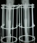
Causes
An anatomical short leg is due to several orthopedic or medical condition mechanisms. Often, one leg simply stops growing before the other one does and is called ?congenital?. We often see mother-daughters or father-sons who exhibit virtually the same degree of shortness on the same side. Often it is not known why this occurs, but it seems to account for approximately 25% of the population demonstrating a true LLD. Other causes of a true LLD include trauma or broken bones, surgical repair, joint replacement, radiation exposure, tumors or Legg-Calves-Perthes disease.
Symptoms
The patient/athlete may present with an altered gait (such as limping) and/or scoliosis and/or low back pain. Lower extremity disorders are possibly associated with LLD, some of these are increased hip pain and degeneration (especially involving the long leg). Increased risk of: knee injury, ITB syndrome, pronation and plantar fascitis, asymmetrical strength in lower extremity. Increased disc or vertebral degeneration. Symptoms vary between patients, some patients may complain of just headaches.
Diagnosis
On standing examination one iliac crest may be higher/lower than the other. However a physiotherapist, osteopath or chiropractor will examine the LLD in prone or supine position and measure it, confirming the diagnosis of structural (or functional) LLD. The LLD should be measured using bony fixed points. X-Ray should be taken in a standing position. The osteopath, physiotherapist or chiropractor will look at femoral head & acetabulum, knee joints, ankle joints.
Non Surgical Treatment
Treatment depends on the amount and cause of the leg length discrepancy as well as the age of your child. Typically, if the difference is less than 2 cm we don?t recommend immediate treatment. We may recommend that your child wear a heel lift in one shoe to make walking and running more comfortable. If the leg length discrepancy is more significant, your doctor may recommend surgery to shorten or lengthen a leg. The procedure used most often to shorten a leg is called epiphysiodesis.

Surgical Treatment
Large leg length inequalities can be treated by staged lengthenings or by simultaneous ipsilateral femoral and tibial lengthenings. Additionally, lengthenings can be combined with appropriately timed epiphysiodesis in an effort to produce leg length equality. Staged lengthenings are often used for congenital deficiencies such as fibular hemimelia, in which 15 cm or more may be needed to produce leg length equality. We typically plan for the final lengthening to be completed by age 13 or 14 years, and allow at least 3 years between lengthenings. Lengthening of both the tibia and femur simultaneously requires aggressive therapy and treatment of soft tissue contractures. Curran et al[57] reported the need for surgical release of soft tissue contractures in 3 of 8 patients treated with simultaneous ipsilateral femoral and tibial lengthenings. Lengthening over an IM nail can be done in an effort to decrease the amount of time the fixator needs to be worn and to prevent angular malalignment. This technique requires that the patient be skeletally mature and it carries a higher risk of osteomyelitis (up to 15%). Additionally, if premature consolidation occurs, a repeat corticotomy is more difficult.
Have you noticed that your pants always fit a little weird or that you are always leaning to one leg when standing for awhile? If so, one of your legs may be longer than the other. This is known as a leg length discrepancy. There are two main reasons for a leg length discrepancy. One reason is that one of your leg bones (tibia or femur) is longer on one side. This is referred to as a true leg length discrepancy because the actual length of your bones is different. A second reason is that your pelvic bone may be rotated on one side making it appear that one leg is longer than the other. This is referred to as an apparent leg length discrepancy because the actual length of your leg bones is not different. In order to figure out if you have a true or apparent leg length discrepancy, your doctor may take an x-ray to measure the length of your leg bones or a simple measurement from your belly button to your ankle can help determine the reason. Over time, the leg length difference can cause stress on your low back, hips and knees, which may cause pain or discomfort.
Causes
An anatomical short leg is due to several orthopedic or medical condition mechanisms. Often, one leg simply stops growing before the other one does and is called ?congenital?. We often see mother-daughters or father-sons who exhibit virtually the same degree of shortness on the same side. Often it is not known why this occurs, but it seems to account for approximately 25% of the population demonstrating a true LLD. Other causes of a true LLD include trauma or broken bones, surgical repair, joint replacement, radiation exposure, tumors or Legg-Calves-Perthes disease.
Symptoms
The patient/athlete may present with an altered gait (such as limping) and/or scoliosis and/or low back pain. Lower extremity disorders are possibly associated with LLD, some of these are increased hip pain and degeneration (especially involving the long leg). Increased risk of: knee injury, ITB syndrome, pronation and plantar fascitis, asymmetrical strength in lower extremity. Increased disc or vertebral degeneration. Symptoms vary between patients, some patients may complain of just headaches.
Diagnosis
On standing examination one iliac crest may be higher/lower than the other. However a physiotherapist, osteopath or chiropractor will examine the LLD in prone or supine position and measure it, confirming the diagnosis of structural (or functional) LLD. The LLD should be measured using bony fixed points. X-Ray should be taken in a standing position. The osteopath, physiotherapist or chiropractor will look at femoral head & acetabulum, knee joints, ankle joints.
Non Surgical Treatment
Treatment depends on the amount and cause of the leg length discrepancy as well as the age of your child. Typically, if the difference is less than 2 cm we don?t recommend immediate treatment. We may recommend that your child wear a heel lift in one shoe to make walking and running more comfortable. If the leg length discrepancy is more significant, your doctor may recommend surgery to shorten or lengthen a leg. The procedure used most often to shorten a leg is called epiphysiodesis.
Surgical Treatment
Large leg length inequalities can be treated by staged lengthenings or by simultaneous ipsilateral femoral and tibial lengthenings. Additionally, lengthenings can be combined with appropriately timed epiphysiodesis in an effort to produce leg length equality. Staged lengthenings are often used for congenital deficiencies such as fibular hemimelia, in which 15 cm or more may be needed to produce leg length equality. We typically plan for the final lengthening to be completed by age 13 or 14 years, and allow at least 3 years between lengthenings. Lengthening of both the tibia and femur simultaneously requires aggressive therapy and treatment of soft tissue contractures. Curran et al[57] reported the need for surgical release of soft tissue contractures in 3 of 8 patients treated with simultaneous ipsilateral femoral and tibial lengthenings. Lengthening over an IM nail can be done in an effort to decrease the amount of time the fixator needs to be worn and to prevent angular malalignment. This technique requires that the patient be skeletally mature and it carries a higher risk of osteomyelitis (up to 15%). Additionally, if premature consolidation occurs, a repeat corticotomy is more difficult.